
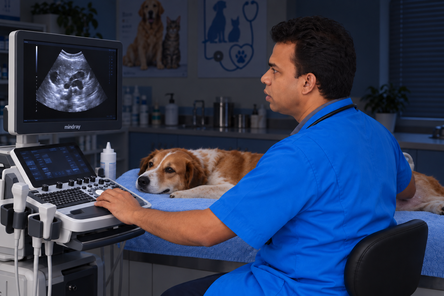
What is an abdominal ultrasound?
The scanner uses sound waves and their echoes to create moving images. It does not use the ionising radiation used for X-rays. The vet can examine organs such as the liver, kidneys, spleen and urinary bladder, as well as parts of the digestive tract. An abdominal examination is different from echocardiography, which focuses on the heart and requires its own views and measurements.
Why has my vet referred my pet?
Reasons may include persistent vomiting, abdominal discomfort, urinary problems, unexplained blood-test changes, or a suspected lump or fluid collection. Ultrasound can also help with selected reproductive questions. A referral is a request for more information: for example, a blood result may suggest an organ problem without showing its structure. Ask which question the scan is intended to answer and whether your pet needs any other care while waiting.
What can the scan tell us—and what can it miss?
Images can reveal changes in organ size, shape or internal appearance. However, gas can obscure parts of the abdomen, and bone blocks the ultrasound beam. Some diseases have similar appearances, and some cause little visible change. A normal scan does not exclude every illness. An abnormal area is not automatically cancer; blood tests, repeat imaging or a sample may be needed to work out what it represents.
Before travelling to the appointment
- Ask the clinic for your pet’s own food, water and medicine instructions. Fasting may be requested for an abdominal scan, but do not apply a general online fasting time to a puppy, kitten, diabetic or unwell animal.
- Ask whether a fuller bladder would help and what to do before arrival. Never force a pet to hold urine when they are uncomfortable or struggling to pass it.
- Bring the referral, earlier blood results and images, medicine names and doses, and a brief symptom timeline. Confirm whether the clinic already has these records.
- For travel from another area, confirm the appointment, expected visit length, estimated charges and report arrangements. Use a secure carrier for cats and a suitable lead for dogs.
What happens during the scan?
The team will review the history and assess your pet. Fur is usually clipped over the area being examined, and gel helps the probe contact the skin. Your pet may lie on their side or back with support. The probe itself does not involve an injection, although pressure over a sore abdomen may be uncomfortable. Tell the team about pain or handling difficulties. Some pets cooperate awake; others need a planned sedative for comfort and useful images.
Is a needle sample part of every ultrasound?
No. A needle sample or biopsy is a separate procedure that may be proposed if an image alone cannot answer the clinical question. Ask what will be sampled, how the result could change treatment, and about bleeding risk, sedation or anaesthesia, laboratory charges and consent. The vet may recommend safety checks first. Do not assume that agreeing to an imaging appointment automatically includes every possible sampling procedure.
Results and the journey home
Ask who will explain the findings and when the report will reach your referring vet. Some answers may be available at the visit, while laboratory results or specialist interpretation can take longer. Confirm when food and usual medicines can resume. If sedation or sampling was performed, obtain the specific supervision and aftercare instructions before leaving. A worsening pet should be reassessed rather than waiting solely for the written report.
Questions owners often ask
- Will every scan find the cause? No. It may narrow the possibilities and guide the next test.
- Can I stay with my pet? Ask the team; handling needs, facilities and any procedure being performed affect this.
- Should I wait for the booking if my pet deteriorates? No. Collapse, severe pain, breathing difficulty or inability to pass urine needs urgent advice and assessment.
General education for dogs and cats; not a diagnosis or an individual treatment plan.